5 | Procedure for Using a Compound Microscope

5 | Procedure for Using a Compound Microscope
Procedure for Using a Compound Microscope
- Compound microscopes can be challenging to use at first and require a bit of practice to produce clear images. It all starts with a well prepared specimen. By following the procedure outlined below, you will soon be getting great results.
- Cut a very thin slice of the sample to be viewed. Cut a very thin slice of the sample to be viewed. Cut a very thin slice of the sample to be viewed. Cut a very thin slice of the sample to be viewed. Cut a very thin slice of the sample to be viewed.
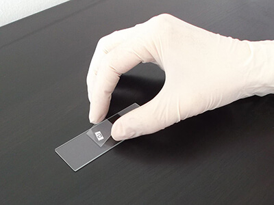
- Place the specimen on a microscope slide.
- Place one or two drops of water on the specimen.
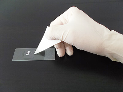
- Gently place a cover slip on the specimen, lowering one side first, then the other. Use a piece of tissue or blotting paper to absorb any excess water that may have spilled out from under the cover slip.
- Place the slide on the stage and secure it in place with the stage clips.
- Select the objective lens with the lowest magnification by rotating the nosepiece.
- Turn on the lamp.
- Use the stage controls to move the slide so that the specimen is directly under the objective lens.
- Adjust the coarse focus knob until the specimen comes into focus.
- * The coarse focus moves the stage up and down. Be very careful not to move the stage up too high that it collides with the objective lens.
- Adjust the fine focus knob to optimise the focus.
- Select a higher magnification objective lens if desired. You may have to readjust the fine focus.
Preparing a good specimen slide takes a bit of practice.
(Images: Witia, Wikimedia Commons)